Neuroscience Microscopy Core
Core Director: Arvydas Matiukas, PhD
Phone: 315 464-7997
Location: 3607 IHP/NRB
Email: [email protected]
这个核心设备使神经科学家能够可视化单个神经元的结构及其在生物体中的活动,并在许多其他研究领域(如微生物学)具有潜在的应用, molecular biology, ophthalmology, etc.) for the study of 3D structure and process dynamics.
The Core provides instruments to image fixed samples and live cells. 硬件和软件允许从单片到多色3D时间序列的不同成像模式, and large area tiling. 可用的环境室为活细胞长期实验提供了最佳条件.
Project consultation/collaboration, training, 核心总监(蔡司认证的显微镜专家)提供技术支持。. 所有用户在操作设备前必须接受培训(每个显微镜单独使用). 经过培训和熟练认证的用户可以在没有监督的情况下操作核心设备.
Instrumentation in Core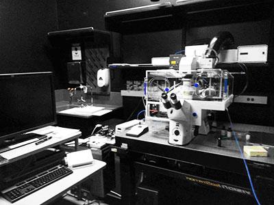
Zeiss LSM 780 confocal reservation calendar
The LSM780 is an inverted microscope with 4 lasers, 3 detectors, an environmental chamber, and mercury and halogen light sources. The microscope has 6 excitation color capabilities, 3个独立的荧光成像通道,包括一个光谱通道, and one transmission PMT. 该系统可配置为同时和/或顺序多色采集.
Leica SP8 with STED reservation calendar

Confocal Microscopy
徕卡的SP8共聚焦系统有五个光谱探测器通道:两个pmt和三个HyDs,以及HyVolution软件,将超灵敏的HyD检测与SVI惠更斯反褶积相结合, 与标准共聚焦显微镜的200-250 nm相比,允许横向分辨率降至140 nm. 激发由405nm激光器和白光激光器(470-670nm连续光谱)提供, up to 8 lines may be combined).
STED (stimulated emission depletion)
3X nanoscopy 是克服衍射极限分辨率的超分辨率技术吗. With multiple STED laser lines of at 592 nm, 660 nm and the pulsed laser at 775 nm, STED can reach a resolution below 30 nm.
Digital light sheet (DLS) Microscopy (currently not online)
DLS显微镜是一种荧光技术,它利用光的平面进行三维成像,这对像胚胎这样的大样本很有用, sensitive samples, or fast biological processes in vivo. 它提供了良好的光学切片能力和高速度与低水平的光漂白.
Deconvolution workstation with Huygens software
惠更斯软件恢复2D和3D显微镜图像或时间序列可视化. The restoration is based on different deconvolution algorithms, 这允许从其共聚焦图像中恢复原始物体/结构,这些图像因模糊和噪声而退化. 反褶积显著提高了图像对比度和分辨率(Z方向约为2x, Z方向约为1.5x in XY).
Facility Rules
If you fail to show up for time you have scheduled on the calendar, you will be billed for the amount of time scheduled
删除(取消)或修改日历上的时间安排必须在您计划的时间之前至少24小时完成 or you will be billed for the amount of time originally scheduled.
New user enrollment:
- The new user downloads and completes the Enrollment Form, emails it to Arvydas.
- Staff coordinates and schedules training date and time.
- More training sessions are scheduled as needed to complete training.
- 新用户被授予与其培训相对应的状态和访问权限.
- 一定要记得在您的出版物中使用Core显微镜和/或工作人员协助生成图像/数据.
Usage:
- 所有使用者在使用显微镜之前必须经过培训. 培训结束后,将建立一个账户,用于预留时间和使用显微镜.
- 所有用户必须在网上日历上注册,以便在使用显微镜之前预留时间, and record actual usage in paper log.
- 使用者必须清洁使用过的显微镜物镜,用完后处理垃圾. 如果您携带任何活体标本或潜在有害样品,请将其处理在您的实验室.
- 请在您的课程结束时查看在线预订日历,以便在最后一刻更改和更新时间表. Leave the system on if someone is reserved next within two hours; keep the lasers in standby whenever possible to prolong their life.
- 晚上的最后一个用户负责关闭显微镜系统.
- You are responsible for your data. The system computers are for temporary image storage. 请将您的数据保存在计算机的数据硬盘上,并在获取后尽快传输您的图像数据. 留在电脑硬盘上的映像文件将定期删除.
- 使用者在使用显微镜时,如有任何问题或损坏,请及时报告.
- No food or drink is allowed in the microscope rooms.
Billing:
- The hours billed are based on your computer log off. Remember to log off to end the billing session.
- All users must record system usage on the paper log at each microscope.
- Minimum time billed is 1 hr
- 每次成像将按预留时间或以1小时为单位使用的小时数中较大者计费.e., all time is round up to a full hour)
- 在前6个小时的培训后,您将收取培训和激光使用的费用
- 技术支持以1分钟为单位计费,并添加到激光使用的成本/小时中
- There is a quarterly cap on the cost of laser use. Other fees (training, 技术支持等)除激光使用外还收费,不打折.
- If you fail to show up for time you have scheduled on the calendar, 您将按照预定的时间付费(这不被认为是激光使用,也不包括在季度折扣中)。.
删除(取消)或修改日历上的时间安排必须在您计划的时间之前至少24小时完成 or you will be billed for the amount of time originally scheduled. (注意日历保留所有条目和修改的电子记录).
Calendar Sign up:
- All calendar reservations are in 1 hr blocks
- Advance reservation is limited:
-
- 从每周五开始,只有下周的时间可以预订
- 每个工作日/实验室的预约时间限制为5小时,应该适合上午8点至下午1点或下午1点至6点的窗口(以1小时为单位)。
- If time is available, you may sign up in the afternoon for additional time, only for the next day (in 1 hr blocks).
- Time reserved on weekends and off hours is not limited in duration, but you may only sign up (starting Friday) for the following week.
- 如果提前完成,请在关闭激光器前联系下一个用户.
- Specific for the Leica - when signing up on the calendar specify the modality you will use. If you need specific objectives contact the core director ([email protected]) when you sign up.
Videos and resources
Videos/resources on fluorescence microscopy and confocal microscopy:
Good Resources
JoVE (wide field, basics of fluorescence microscopy of samples)http://www.jove.com/v/5040/introduction-to-fluorescence-microscopy?list=UicAJpg9
Microcourses from Harvard
http://www.youtube.com/channel/UC4cOKa0TZK8CQhzSQqfwuMQ
iBiology Microscopy courses
http://www.ibiology.org/online-biology-courses/microscopy-series/
Recommended for fluorescence microscopy and confocal overviews
http://www.jove.com/v/5040/introduction-to-fluorescence-microscopy?list=UicAJpg9
http://www.youtube.com/watch?v=9o5E27dnauk&list=PL7Y2NBzyw8AhVl5iJZIACsq-Rk8fmB5f9&index=4
http://www.youtube.com/watch?v=mGPJBNsq7bA&list=PL7Y2NBzyw8AhVl5iJZIACsq-Rk8fmB5f9&index=5
Preventing Damage
Preventing lens damage:Preventing Objective Lens Damage: Blunt Force Trauma
Immersion Oil Problems
http://youtu.be/c58P4Zt9xX0?list=PL7Y2NBzyw8AioI3ea7en6jOZXTTrlxVF2
Microscopy Course on iBiology
Popular, free online microscopy course begins with basics of optics, proceeds through transmitted light microscopy, and covers many microscopy methods.http://www.ibiology.org/talks/fluorescence-microscopy/
http://www.ibiology.org/talks/confocal-microscopy/
More Information
SUNY Upstate Medical University and Leica Microsystems, Inc. 共同努力在纽约州立大学推荐最近最火的赌博软件建立徕卡微系统卓越中心. 该中心支持一项使命,即利用最先进的成像系统,推动科学研究的新发现和新见解.
- LAS X Software introduction and download: http://go.leica-ms.com/SUNYLASX
- STED microscope manual: http://go.leica-ms.com/SUNYSTED3X
- DLS microscope introduction: http://go.leica-ms.com/SUNYDLS
- Leica Microsystems Tutorials: http://www.youtube.com/channel/UCLBTBR4a0IR64CL6X9wjbwQ/videos